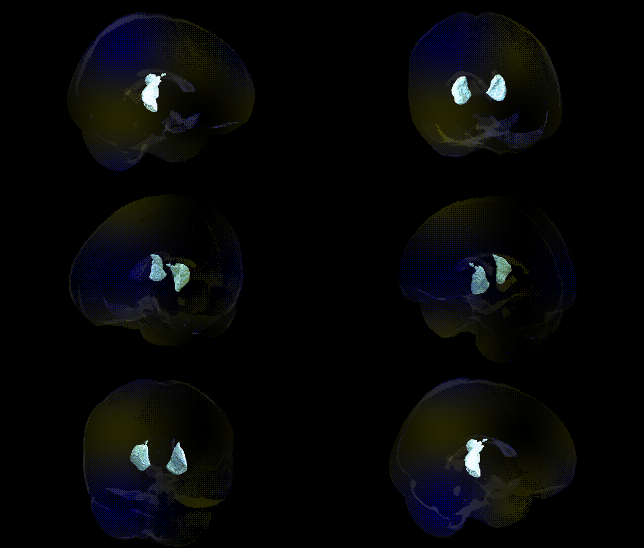
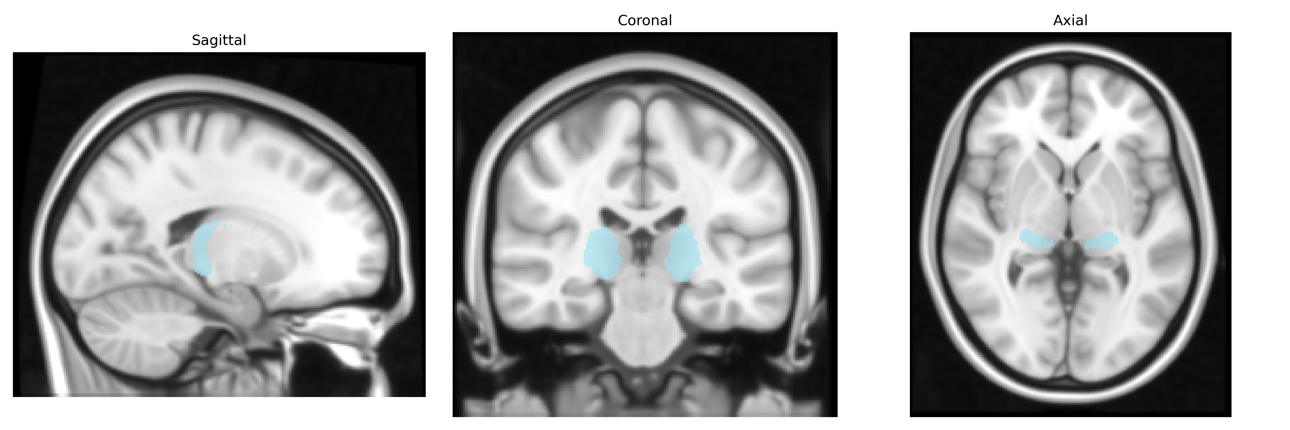
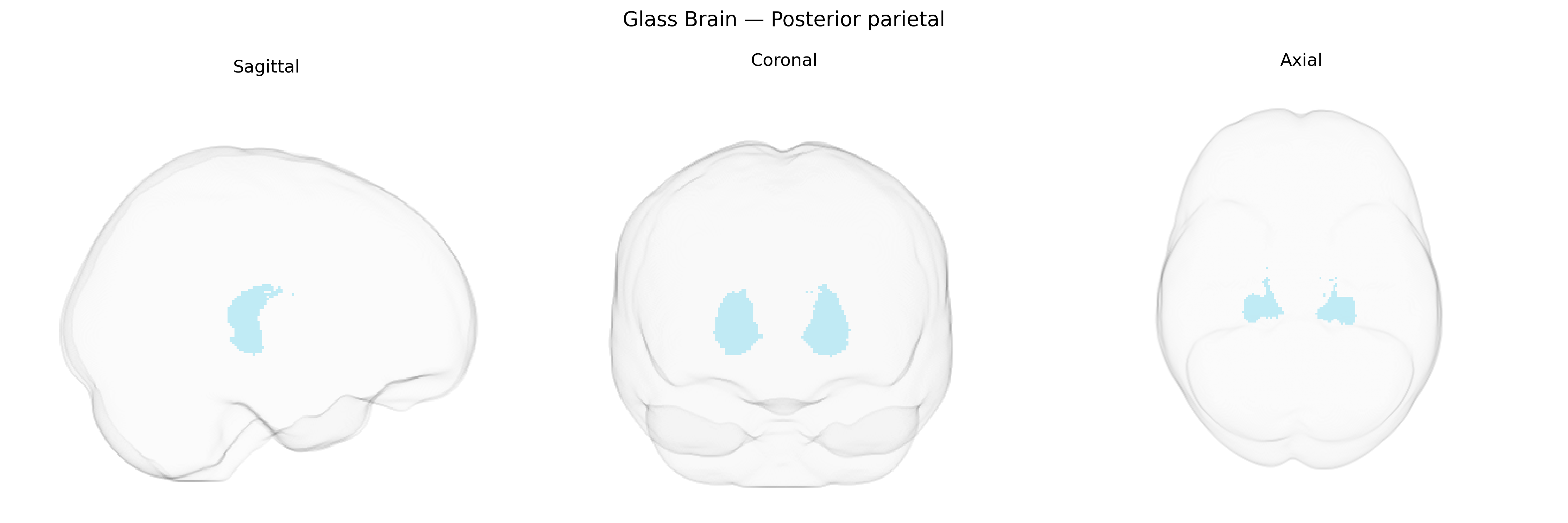
The bilateral Posterior parietal region in the Thalamus maxprob thr25 1mm Atlas corresponds to thalamic nuclei projecting predominantly to the posterior parietal cortex, a multimodal association area involved in visuospatial integration, attention, sensorimotor transformation, and higher-order perception of body and extrapersonal space. Functionally, this thalamic group relays and integrates inputs from sensory systems (particularly visual and somatosensory) and subcortical motor structures, contributing to the formation of spatial maps and the coordination of goal-directed movements, including reaching and eye–hand coordination. Through reciprocal connections with parietal association cortex and other cortical regions, it participates in the network supporting spatial attention, perception of motion and position, and the transformation of sensory signals into motor plans. There is no direct Wikipedia article for this specific thalamic “Posterior parietal” label; a closely related cortical target region is the Posterior parietal cortex.
Genetic associations involving the bilateral posterior parietal region, as defined in thalamocortical and whole-brain atlases such as the Thalamus maxprob thr25 1 mm, largely derive from GWAS and imaging-genetics studies linking parietal structure and function to polygenic influences on cognition and neuropsychiatric risk. Variants in genes related to synaptic plasticity, neuronal migration, and axon guidance (for example, BDNF, DISC1, NRG1, and genes in calcium and glutamatergic signaling pathways) have been associated with altered parietal cortical thickness, surface area, or functional activation, and these same variants often contribute to risk for schizophrenia, autism spectrum disorder, ADHD, and major depressive disorder, where posterior parietal abnormalities are frequently reported. Large-scale GWAS of brain imaging phenotypes (ENIGMA, UK Biobank) have identified loci in and around genes such as HMGA2, IGF1, FGFR3, and those involved in Wnt and MAPK signaling that influence parietal lobe morphology and white-matter connectivity, including thalamoparietal tracts, with downstream effects on general cognitive ability, attention, working memory, and educational attainment. In addition, polygenic scores for Alzheimer’s disease, frontotemporal dementia, and other neurodegenerative conditions show associations with posterior parietal atrophy and hypometabolism, complementing earlier candidate-gene work on APOE and related lipid-processing genes. Overall, the genetic architecture of the bilateral posterior parietal region appears highly polygenic and overlapping with that of multiple cognitive traits and neuropsychiatric disorders, with many implicated variants exerting broad, distributed effects rather than region-specific influences.
Overview generated by GPT-4o (2026).
Region ID: 6
Hemisphere: bilateral
Atlas: Thalamus maxprob thr25 1mm
Full Quality Version: Download MP4
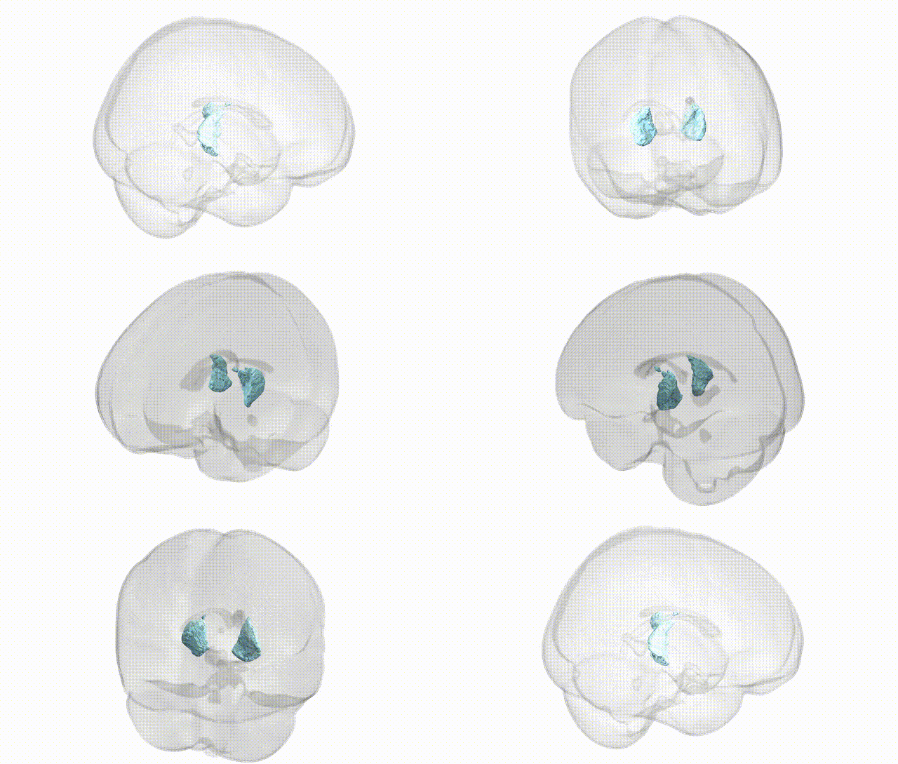
Full Quality Version: Download MP4
Wali Sidiqyar*, Gaurav Rudravaram*, Elyssa M. McMaster, Trent M. Schwartz, Adam M. Saunders, Kurt G. Schilling, Bennett A. Landman "Introducing SPINS: A Shared Public Visualization Library of Neuroanatomical Structures." Medical Imaging with Deep Learning- short paper
This resource is licensed under CC0 1.0 Universal (Public Domain).